MRI ordering is a critical step in diagnostic imaging‚ ensuring appropriate use of magnetic resonance imaging. This guide helps healthcare providers optimize MRI requests for accurate patient care.
1.1 Overview of MRI and Its Importance
Magnetic Resonance Imaging (MRI) is a non-invasive diagnostic tool that provides detailed images of internal body structures. It uses magnetic fields and radio waves to visualize organs‚ tissues‚ and abnormalities. MRI is particularly valuable for soft tissue evaluation‚ such as the brain‚ spine‚ and joints. Its high-resolution imaging helps diagnose conditions like tumors‚ injuries‚ and neurological disorders. Unlike CT scans‚ MRI avoids ionizing radiation‚ making it safer for long-term use. This technology is critical for early detection‚ treatment planning‚ and monitoring disease progression‚ enhancing patient outcomes and clinical decision-making.
1.2 Purpose of an MRI Ordering Guide
An MRI Ordering Guide serves as a valuable resource for healthcare providers to optimize the use of MRI imaging. It helps ensure that exams are appropriate‚ cost-effective‚ and aligned with clinical best practices. The guide provides clarity on when and how to order MRIs‚ reducing unnecessary scans and improving patient care. It also addresses patient preparation‚ safety considerations‚ and interpretation of results. By streamlining the ordering process‚ the guide enhances diagnostic accuracy‚ reduces costs‚ and improves overall healthcare outcomes. It is an essential tool for making informed‚ evidence-based decisions in radiology.
Key Considerations Before Ordering an MRI
Before ordering an MRI‚ consider the clinical justification‚ patient safety‚ and cost-effectiveness. Ensure the exam aligns with evidence-based guidelines and addresses the patient’s specific condition effectively.
2.1 Clinical Indications for MRI
MRI is indicated for evaluating soft tissue injuries‚ joint disorders‚ and neurological conditions. It excels in imaging brain‚ spinal cord‚ and musculoskeletal systems. Use it for stroke diagnosis‚ tumor detection‚ and assessing ligament or tendon damage. MRI is also valuable for non-invasive cardiovascular imaging and detecting abnormalities in organs like the liver and pancreas. Always align the MRI request with the patient’s symptoms and clinical suspicion to ensure optimal diagnostic yield.
2.2 Patient History and Pre-Exam Preparation
Obtaining a detailed patient history is crucial before ordering an MRI. This includes reviewing medical history‚ allergies‚ and previous surgeries. Patients must disclose any metal implants or claustrophobia. Pre-exam preparation involves removing metallic items and ensuring appropriate clothing. Fasting may be required for abdominal MRI. Inform patients about the procedure duration and noise. Provide clear instructions on medications and contrast agents if needed. Ensuring patient compliance with preparation guidelines enhances safety and image quality‚ leading to accurate diagnostic results.

How to Choose the Right MRI
Selecting the appropriate MRI modality depends on the patient’s condition‚ anatomical region‚ and desired imaging detail. Consider factors like magnetic field strength and specialized techniques.
3.1 Types of MRI Modalities (e.g.‚ 1.5T vs. 3T)
MRI modalities vary by magnetic field strength‚ with 1.5T and 3T being the most common. A 3T MRI offers higher resolution and faster imaging but may not be suitable for all patients due to stronger magnetic fields. Open or wide-bore systems are ideal for claustrophobic or larger patients‚ while closed systems provide higher image clarity. The choice depends on the anatomical region‚ clinical indication‚ and patient tolerance. Understanding these differences helps in selecting the most appropriate modality for accurate diagnosis and optimal patient comfort.
3.2 Specialized MRI Techniques (e.g.‚ Contrast-Enhanced‚ Functional MRI)
Specialized MRI techniques enhance diagnostic capabilities. Contrast-enhanced MRI uses gadolinium-based agents to highlight tissue abnormalities‚ improving lesion detection. Functional MRI (fMRI) measures brain activity‚ useful for neurological disorders. Diffusion-weighted imaging (DWI) is crucial for stroke assessment‚ while magnetic resonance angiography (MRA) visualizes blood vessels. Each technique offers unique benefits‚ such as better visualization or functional insights‚ aiding in precise diagnosis and treatment planning. Selecting the right technique depends on the patient’s condition and the clinical question‚ ensuring targeted and effective imaging.

Patient Preparation for an MRI
Patient preparation involves removing metal objects‚ changing into appropriate clothing‚ and fasting if required. Arrive early‚ inform staff of claustrophobia‚ and follow specific instructions provided.
4.1 Provider Responsibilities
Providers must ensure patients are medically suitable for MRI by reviewing their history for contraindications like metal implants or claustrophobia. They should explain the procedure‚ duration‚ and expectations‚ providing clear instructions on removing metal objects and wearing appropriate clothing. Patients should be advised to arrive early and disclose any anxiety. Providers are also responsible for addressing questions‚ ensuring compliance with safety protocols‚ and confirming necessary preparations like fasting if required. Effective communication ensures a smooth and safe MRI experience tailored to individual patient needs.
4.2 Patient Instructions and Education
Patient education is crucial for a successful MRI. Patients should be instructed to remove all metal objects‚ including jewelry and glasses‚ and wear loose‚ metal-free clothing. They must arrive 30 minutes early to complete paperwork and undress if needed. Clear instructions on fasting‚ if required‚ should be provided. Emphasize the importance of remaining still during the scan and inform them about potential claustrophobia management options. Encourage patients to ask questions and ensure they understand the procedure’s purpose and duration to reduce anxiety and ensure cooperation during the exam.
Ordering and Scheduling an MRI
Ordering an MRI requires following specific guidelines‚ selecting the appropriate modality‚ and ensuring clinical justification. Clear communication between providers and radiology departments is essential for accurate scheduling.
5.1 Steps to Order an MRI
To order an MRI‚ begin by identifying the clinical indication and selecting the appropriate modality. Prepare a detailed order with patient information‚ medical history‚ and specific imaging requirements. Use standardized templates or electronic health records (EHR) to ensure clarity. Include relevant prior studies or comparison exams. Verify insurance coverage and obtain pre-authorization if required. Submit the order through the radiology information system (RIS) or directly to the imaging center. Confirm patient availability and schedule the exam at a convenient time‚ ensuring proper preparation instructions are provided.
5.2 Scheduling and Timing Considerations
When scheduling an MRI‚ ensure the imaging center has availability and the necessary equipment. Prioritize urgent cases‚ such as emergencies or time-sensitive diagnoses. Consider the patient’s schedule and medical condition to avoid delays. Provide clear instructions on preparation‚ such as fasting or medication restrictions. Confirm the exam duration to manage patient expectations. Ensure timely communication between the ordering provider and the imaging center to address any issues. Proper scheduling minimizes wait times and ensures efficient patient care while optimizing resource utilization.

Understanding MRI Results
Accurate interpretation of MRI results by radiologists is crucial for diagnosis. Clear communication of findings ensures clinicians can make informed decisions‚ impacting patient care and treatment planning effectively.
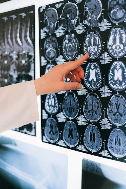
6.1 Interpreting MRI Reports
An MRI report includes clinical history‚ findings‚ and impressions. Radiologists highlight abnormalities‚ such as tissue damage or tumors‚ and suggest further diagnostic steps. The report’s structure helps clinicians quickly identify key information. Understanding terms like T1 and T2 weighted images is essential for interpreting results accurately. Clear communication of findings ensures proper patient care. Any ambiguity in the report should be clarified with the radiologist to avoid misunderstandings or unnecessary additional tests. Advanced techniques‚ like contrast-enhanced MRI‚ may provide more detailed insights‚ aiding in precise diagnosis and treatment planning.
6.2 Communication Between Radiologists and Clinicians
Effective communication between radiologists and clinicians is vital for accurate interpretation and appropriate patient care. Radiologists provide detailed reports‚ while clinicians offer clinical context‚ ensuring comprehensive understanding. Standardized terminology and clear reporting formats enhance collaboration. Regular feedback loops help clarify ambiguities and improve future reporting. Both parties should actively engage in discussions to align findings with clinical suspicions‚ optimizing diagnostic accuracy. This collaborative approach ensures that MRI results are translated into actionable insights‚ directly benefiting patient outcomes and treatment planning.

Safety and Contraindications
MRI safety requires screening for metal objects‚ claustrophobia‚ and medical implants. Contraindications include pacemakers‚ aneurysm clips‚ and certain implants. Pregnancy and kidney issues may also pose risks.
7.1 MRI Safety Guidelines
MRI safety guidelines are essential to ensure patient and staff protection. Screening for metal objects‚ claustrophobia‚ and medical implants is critical. Ferromagnetic materials pose risks‚ requiring thorough patient evaluation. Claustrophobia may necessitate sedation or open MRI use. Implantable devices‚ such as pacemakers or aneurysm clips‚ must be verified for MRI compatibility. Pregnancy and kidney function are also considerations‚ as gadolinium contrast agents may be contraindicated in severe kidney disease. Adhering to these guidelines minimizes risks and ensures safe imaging procedures.
7.2 Contraindications for MRI
Contraindications for MRI include certain medical implants like traditional pacemakers and aneurysm clips. Ferromagnetic foreign bodies‚ such as shrapnel‚ pose significant risks. Severe claustrophobia without sedation or open MRI access is another contraindication. Renal failure patients with GFR below 30 mL/min/1.73m² may be unable to receive gadolinium-based contrast due to fibrosis risks. Pregnant women‚ especially in early stages‚ should avoid MRI unless medically necessary. These contraindications ensure patient safety and prevent complications during imaging.
Cost Considerations and Insurance
MRI scans can be costly‚ with prices varying by location and modality. Insurance coverage differs‚ often requiring pre-authorization for non-emergency scans.
8.1 Understanding Costs Associated with MRI
The cost of an MRI varies based on factors like location‚ type of scanner‚ and whether contrast is used. On average‚ MRI scans can range from $700 to $2‚000 or more.
8.2 Insurance Coverage and Billing
Insurance coverage for MRI scans varies by provider and policy. Most plans cover MRI when deemed medically necessary‚ though patient out-of-pocket costs may apply. Billing processes involve submitting claims to insurance‚ with providers handling the paperwork. Patients should verify coverage details beforehand to understand potential costs. Additionally‚ some imaging centers offer financing options or discounted rates for uninsured individuals‚ ensuring accessibility to necessary diagnostics.
9.1 Summary of Key Points
9.2 Future Directions in MRI Technology
Advancements in MRI technology are expected to enhance diagnostic capabilities significantly. Higher magnetic field strengths‚ such as 7T scanners‚ will improve image resolution and detection of subtle abnormalities. Artificial intelligence will play a larger role in automating image analysis and reducing interpretation time. Functional MRI (fMRI) will continue to advance‚ offering deeper insights into brain function and neurological disorders. Portable MRI systems may become more accessible‚ enabling imaging in remote or emergency settings. These innovations promise to make MRI more efficient‚ accessible‚ and valuable for both clinical practice and research.